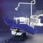
Dental X-ray equipment is a cornerstone of modern dentistry, enabling practitioners to diagnose and treat oral health issues with remarkable precision. However, the use of ionizing radiation in these devices necessitates a thorough understanding of safety protocols, maintenance requirements, and best practices. This guide aims to clarify the complexities surrounding dental X-ray equipment, offering valuable insights for both dental professionals and patients.
Understanding Dental X-Ray Technology
Dental X-ray machines utilize low levels of radiation to capture detailed images of the teeth, gums, and surrounding structures. These images, known as radiographs, are essential for identifying issues that may not be visible during a routine examination. Common conditions detected through dental X-rays include:
- Cavities between teeth
- Bone loss associated with gum disease
- Abscesses or cysts
- Tumors
- Impacted teeth
- Developmental abnormalities
Types of Dental X-Ray Equipment
Different types of dental X-ray equipment serve specific diagnostic purposes:
- Intraoral X-rays: The most common type taken inside the mouth, including:
- Bitewing X-rays
- Periapical X-rays
- Occlusal X-rays
- Extraoral X-rays: Taken outside the mouth to provide a broader view of the jaw and skull. Examples include:
- Panoramic X-rays
- Cephalometric projections
- Cone Beam Computed Tomography (CBCT)
- Handheld X-ray devices: Portable units that offer flexibility in various clinical settings.
Key Safety Considerations for Dental X-Ray Equipment
While dental X-rays involve exposure to ionizing radiation, the levels are generally low and considered safe when proper precautions are taken. Here are essential safety considerations:
Radiation Protection Principles
- Justification: Only take X-rays when there is a clear clinical need.
- Optimization: Apply the ALARA principle (As Low As Reasonably Achievable) to minimize exposure.
- Dose Limitation: Adhere to recommended dose limits for both patients and staff.
Patient Safety Measures
- Use lead aprons and thyroid collars to shield patients from scattered radiation.
- Implement digital radiography to reduce radiation exposure compared to traditional film-based systems.
- Adjust exposure settings based on patient size and the area being examined.
- Follow frequency guidelines for dental X-rays according to patient age and risk factors.
Operator Safety Protocols
- Maintain a safe distance from the X-ray source during exposure, ideally behind a protective barrier.
- Wear personal dosimeters to monitor cumulative radiation exposure.
- Use lead aprons if required to be in the room during exposure.
- Regularly check and maintain equipment to prevent radiation leakage.
Maintenance of Dental X-Ray Equipment
Proper maintenance of dental X-ray equipment is crucial for ensuring safety, image quality, and longevity. Here’s a comprehensive maintenance plan:
Daily Checks
- Inspect cables and connections for wear or damage.
- Clean and disinfect equipment surfaces according to manufacturer guidelines.
- Listen for unusual noises or odors during operation.
Weekly Maintenance
- Test all safety features, including emergency stop buttons and warning lights.
- Verify the alignment of the X-ray beam using alignment test tools.
- Clean and inspect image receptors (for digital systems).
Monthly Tasks
- Conduct thorough inspections of all components.
- Check and clean cooling systems if applicable.
- Review and update equipment logs and maintenance records.
Annual Service
Schedule comprehensive inspections by qualified technicians to calibrate equipment accurately and replace worn parts as recommended by manufacturers.
Quality Assurance Testing
Regular quality assurance testing is vital:
- Conduct image quality tests to ensure optimal diagnostic value.
- Perform radiation output tests to verify consistency and safety.
Best Practices for Using Dental X-Ray Equipment
Implementing best practices ensures safe and effective use of dental X-ray equipment:
- Proper Training: Ensure all operators receive comprehensive training on equipment use, safety protocols, and radiation protection.
- Patient Selection: Use clinical judgment to determine the necessity of X-rays for each patient based on their oral health history and risk factors.
- Equipment Selection: Choose the appropriate type of X-ray for each diagnostic task to minimize unnecessary radiation exposure.
- Technique Optimization: Use correct exposure settings and positioning techniques for high-quality images with minimal radiation.
- Image Quality Control: Regularly assess image quality and implement corrective measures if needed.
Regulatory Compliance and Guidelines
Dental practices must adhere to various regulations governing the use of X-ray equipment:
- Ionising Radiations Regulations 2017 (IRR17): Covers equipment safety requirements protecting workers and the public.
- Ionising Radiation (Medical Exposure) Regulations 2017 (IRMER17): Focuses on patient protection during medical exposures.
- National and International Standards: Follow guidelines from organizations such as the American Dental Association (ADA) and the European Academy of DentoMaxilloFacial Radiology (EADMFR).
Future Trends in Dental X-Ray Technology
The field of dental radiography is continuously evolving with exciting developments on the horizon:
- Artificial Intelligence (AI) Integration: AI algorithms may assist in image interpretation, reducing human error while improving efficiency.
- 3D Printing from X-ray Data: Advanced imaging techniques could allow for creating 3D models that enhance treatment planning and patient education.
- Radiation-Free Imaging Alternatives: Research into non-ionizing imaging methods may provide safer alternatives in the future.
Conclusion
Dental X-ray equipment is an indispensable tool in modern dentistry that provides essential diagnostic information supporting optimal oral health care. By understanding this technology, implementing robust safety measures, maintaining equipment diligently, and following best practices, dental professionals can maximize the benefits of X-ray imaging while ensuring patient safety.
As technology advances, staying informed about developments in dental radiography is crucial. By embracing a culture of safety and continuous improvement, dental practices can deliver high standards of care while minimizing risks associated with radiation exposure. Remember that safe use of dental X-ray equipment relies on knowledge, vigilance, and adherence to established protocols—qualities that ultimately benefit both patients and practitioners alike.


